Chapter 4
Notes
Epithelial and Connective Tissue
Histology: the study of
tissues
1. Definition
- a tissue is a group of similar cells with
intercellular material that together perform a specific
function.
- Four basic tissues and germ layer
derivative
1. Epithelial tissue: ectoderm,
endoderm, mesoderm
2. Connective tissue: mesoderm
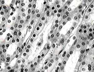
3. Muscle tissue:
mesoderm
4. Nervous tissue:
ectoderm
Epithelial
Tissue
2. Characteristics
- Function - protect, absorb, secrete
- Exist in sheets of cells (membranes)
- No blood supply
- Contain an inferior basement membrane
- anchors epithelial tissue to
connective tissue below
- is composed of connective tissue
fibers and
intercellular
substance
- Types of Epithelial Cells
- Squamous - flat, irregular cells
- Columnar - column-shaped with oval
nucleus at base of cell
- Cuboidal - relatively square;
central nucleus
- Method of Classifying Epithelial Tissue
- by cell type
- by number of cell layers
simple - one cell layer
stratified - multiple cell layers
- Epithelial
tissue online comparison
3. Types of Epithelial Tissue
Simple Squamous epithelium

- Structure - single layer of
flat, squamous cells
- Function - flattened
cells emphasize ability to absorb
- Location - air
sac (alveoli) of lungs, capillary walls,
covering
of organs, part of kidney that filters blood
Simple cuboidal epithelium
- Structure - single layer of cuboidal cells
- Function - absorption, secretion
- Location - kidney tubules,
lines cornea & lens of eye,
Thyroid gland
Simple columnar epithelium
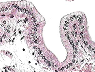
- Structure - single layer of columnar
cells
- may contain the following specialized structures
a. Goblet cells - specialized for mucus secretion
b. Microvilli with perpendicular projections for
absorption
c. Cilia in respiratory tract
- Function - protection, secretion,
absorption
- Location - lining of digestive tract, gall
bladder and part
of uterine tubes & respiratory tract
Keratinized stratified squamous
epithelium
- Structure - multiple layers of three
epithelial cell
types, superficial cells, dehydrate, fuse, and
fill with keratin, a yellow, horny material
- basal layer undergoes mitosis
- Function - protection of living cells
below
- first line of defense
- Location - surfaces constantly exposed to
air; skin
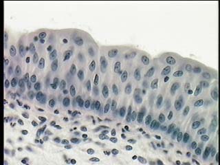
Nonkeratinized stratified squamous
epithelium
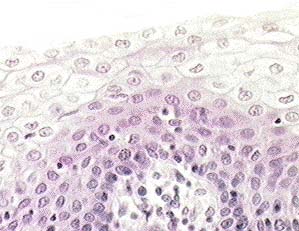
- Structure - multiple layer of three
epithelial cell
types; contains mucous glands
- Function - protection from abrasion
- Location - lining of mouth, esophagus,
vagina
Pseudostratified columnar epithelium
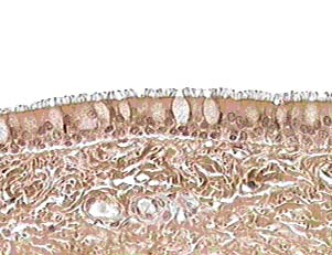
- Structure - single layer of columnar cells
with
appearance of multiple layers
- may contain goblet cells and cilia
- Function - protection, trap dust particles
- Location - lining of trachea,
male reproductive
ducts, ducts of some glands
Transitional
epithelium
- Structure - all 3 types of cells with
rounding of outer layer
- Function - allow for stretch (distension)
- Location - lining of urinary
bladder and ureters
Glandular Epithelium
- Definition - specialized group of
epithelial cells that function in secretion
- Formation of Glands
- epithelial cells grow downward into the supportive
tissue
- cells proliferate and differentiate
- endocrine
glands
- cells that detach from epithelium
- ductless
- secretions
are released into blood stream
- exocrine glands
- cells that
retain their connection with the epithelial surface
- duct is
present
- secretion
released onto surface of epithelium
-types of
exocrine glands
1. Halocrine - cell dies and releases content
- sebaceous gland
2. Merocrine - secretion released without any damage to
cell
- sweat gland, pancreas, salivary
3. Apocrine - secretion pinches off from cell
- mammary gland
Histology
online sites
Connective Tissue
4. Characteristics
- Function - support and binding
- Cells are scattered among fibers and a
matrix
- Vascular
- Arises from embryonic mesenchyme
- Matrix varies from fluid to solid, holds
fibers and cells
in place while determining the function of the tissue
- Fibers are made from three proteins
- Types of fibers
a.
Collagenous - contains the protein collagen,
- fibers are strong and flexible
b. Reticular
fibers - fine branching fibers form
a supporting framework
c. Elastic
fibers - protein is elastin, fibers have
strength and elasticity
- Types of Connective Tissue Cells
a.
Fibroblast - produces fibers
& matrix
- most numerous
- involved in repair and growth
b.
Fibrocyte - mature
fibroblast
- maintenance
c. Macrophages
- defense, phagocytosis
d. Plasma
cells - source of circulating antibodies
e. Mast Cell -
releases heparin, an anti-coagulant
- releases histamine, dilates small blood vessels
f. Fat Cell -
stores triglycerides
- signet ring shape
5. Types of Connective Tissue
Areolar connective tissue (areolar)
- Structure - collagenous &
elastic fibers
- all 6 types of connective tissue cells
- fluid matrix contains
hyaluronic acid
which aids in diffusion
- Function - covers organs
- holds blood vessels & nerves in place
- widely distributed
- nutritive role
- second line of defense
- Location - mucous membranes
- between tissue of body organs
- with adipose tissue forms subcutaneous layer
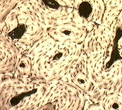
Dense connective tissue
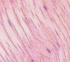
- Structure - collagenous fibers
predominate
- cells are fibroblasts and macrophages
- dense matrix
- Function - provides strength
- Location - fibers arranged in
parallel bundles for strength
- tendons (attaches muscle to bone)
- ligaments (holds bones to joints)
- fibers irregular for stretch aponeuroses (fasciae)
and capsules of organs
Elastic connective tissue
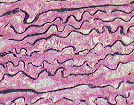
- Structure - free branching elastic
fibers, few fibroblasts
- Function - allows
expansion and recoil
- Location - lungs, trachea, arteries,
aorta
Reticular connective tissue
- Structure - mainly reticular
fibers, thin matrix
- Function - holds cells of
loose organs together
- Location - liver, spleen, bone
marrow
Adipose connective tissue
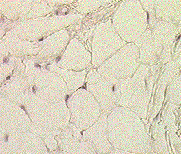
- Structure - collagenous and elastic
fibers
- all six connective tissue cells
- fibroblasts specialize as fat cells
(central fat vacuole thin cytoplasm)
- associated with areolar connective tissue
- Function - food reserve for
energy
- prevents loss of body heat
- Location - around most organs
- beneath skin
- marrow of long bones
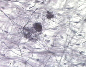
Cartilage
- General characteristics
1. cells are chondrocytes
2. matrix is semisolid containing
chondroitin
3. lacuna - a depression in matrix
which houses chondrocytes
4. perichondrium - connective tissue
membrane around cartilage
5. no blood supply
- Types
Hyaline cartilage
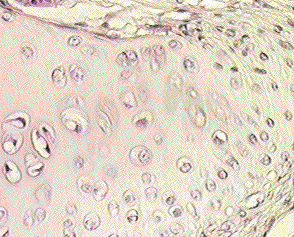
- Structure - invisible fine
collagenous fibers
- two chondrocytes housed in each lacunae
- thick gelatinous ground substance
- Function - acts as a model for
embryonic bone
formation, prevents tissue damage
from friction. Provides shape to nose
and respiratory passages
- Location - covering bone ends at joints
- tip of nose
- between rib and sternum (costal)
- epiphyseal plate
Fibrocartilage
- Structure - collagenous fibers
arranged in parallel bundles
- chondrocytes sandwiched between bundles
- Function - provides strength
- Location - intervertebral
discs, pubic symphysis
Elastic cartilage
- Structure - many elastic fibers
- Function - allows for bending with
return to original shape
- Location - external ear, larynx and
eustachian tubes
Bone tissue
- General Characteristics
- organic
matter - 35% (cells & fibers)
- inorganic
material - 65% (matrix & calcium salts)
- Types of bone cells
1. osteocytes - maintenance of intercellular material
(matrix)
2. osteoblasts - peripheral bone forming cells
3. osteoclasts - internal, actively destroy bone matrix
- Classification of bone according
to structure
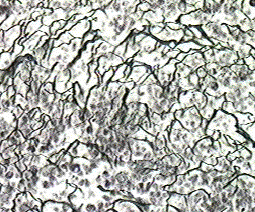
1. Compact
bone
- arranged into concentric rings called Haversian systems
- provides strength
- is external & solid
- Haversian system consists of:
lamella - concentric ring of matrix
lacuna - openings between lamellae for osteocytes
osteocytes - mature bone cell
Haversian canal - in center of lamella; houses vessels
Canaliculi - radiating channels between lacuna
and Haversian canal for nutrients and wastes
Volkmann canal - crosswise canals from Haversian canal
to exterior containing blood vessels and nerves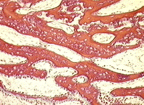
2. Spongy bone
- irregular lattice work of bone called trabecula
- spaces filled with red bone marrow
- osteocytes trapped within calcium matrix
Connective Tissue Lab